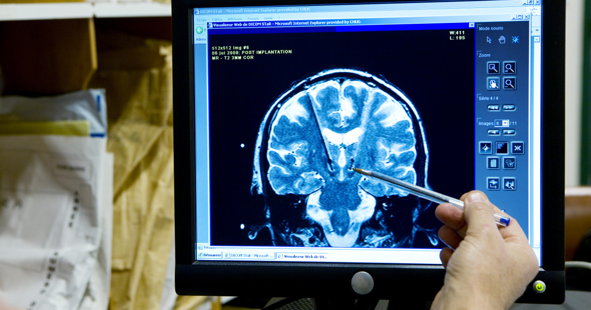
With the help of Google, researchers from Harvard University have created the highest-resolution 3D map ever of the structure of the human brain. Although the map only addresses one cubic millimeter piece of the brain, 1.4 petabytes (1,434 terabytes) of raw data were generated during the description.
This is perhaps the most computationally demanding task in brain research. A daunting task
– said Michael Horelich, an employee of the Allen Institute for Brain Research, as an independent professional.
Current brain atlases have lower resolution, but the point of contact between one neuron and another can be traced back to this nanoscale.
If we really want to understand how the brain processes information and stores memories, we will eventually need a map of that decision
– noted Viren Jain, Senior Researcher at Google.
There are many anatomical maps of the human brain. There is one that depicts the arrangement of neurons, and one that depicts gene expression. As can be seen from the above, this describes nerve connections. A cubic millimeter piece of tissue contains 57,000 cells and 230 millimeters of capillaries, as well as approximately 183 million nerve connection points, or synapses.
The entire brain is a million times larger than that. The cerebral cortex contains about 16 billion neurons, with trillions of connections between them. However, obtaining tissue was not easy, and a deceased donor could not be considered, because this tissue begins to decompose very quickly. Instead, he is 45 years old and undergoing surgery for epilepsy
A patient donated tissue to researchers.
This small sample was prepared using epoxy resin, then cut into 5,019 30-nm slices with a diamond blade, and imaged using a high-speed electron microscope specially developed for this purpose. The slices were then matched and grouped by a machine learning system, and individual nerve fibers were color-coded. There was still a lot of work to be done when the first reports of the research became scientific buzz in 2021.
There are all these cables coming and going everywhere to make all kinds of connections
Jain explained, adding that untangling the situation is more difficult than we think.
If you mess up a cable, all the connections on that cable will be wrong.
The research immediately revealed scientific surprises: the nerve extensions carrying the signals had curled up in some places and formed a structure that revolved around them. Axons, which are similar extensions that terminate at a single synapse, were sometimes involved in multiple connections, with as many as fifty synapses.
You can find a lot of things in it that textbooks don't talk about
noted Jeff Lichtman, a brain researcher at Harvard University.
According to the brain research profession, with this 3D map, we are closer than ever to learning about the internal structure of the brain.
Two decades ago Caenorhabditis elegans meningitis The 302-neuron ringworm nervous system was the first complete brain map, followed by an electroencephalogram of the fruit fly. There are currently research programs called MouseLight and MICrONS, which map smaller parts of the mouse brain. Of course, the human brain is a completely different world. The structures revealed for the 3D map are a Neuroglancer The search can be viewed using a web application called From its website.
(Earth.com, MIT Technology Review, Singularity axis)


